Your heart’s normal, or sinus, rhythm is controlled by a natural pacemaker, the sinus node, which creates electrical impulses that travel across the atria to the ventricles, causing them to contract and pump blood out to your lungs and body in what is known as normal sinus rhythm. Symptoms of PVCs include a fluttering or flip-flop feeling in the chest, pounding or jumping heart rate, skipped beats and palpitations, or an increased awareness of your heartbeat.

PVCs - also called also called premature ventricular complexes, ventricular premature beats and extrasystoles - are very common and usually harmless. While BBR VT is also quite amenable to catheter ablation, it generally occurs in patients with significant structural heart disease (cardiomyopathy) thus most patients with BBR VT also warrant ICD therapy.Premature ventricular contractions (PVCs) are extra, abnormal heartbeats that begin in the ventricles, or lower pumping chambers, and disrupt your regular heart rhythm, sometimes causing you to feel a skipped beat or palpitations. An additional VT-type, bundle branch re-entry (BBR), usually has an appearance indistinguishable from LBBB SVT (propagation anterogradely over RBB, retrogradely over LBB) and thus belongs in the general category of VTs that are difficult (if not impossible) to distinguish from SVT. 21 Diagnosis of these forms of VT is more difficult than the VT-related to structural heart disease, because the QRS duration is comparatively shorter, VA conduction is often present (not dissociated), and many other criteria fail to distinguish these VTs from SVT (as in Figure 4). These largely benign forms of VT are particularly important their presence, in the setting of a structurally normal heart, is a contraindication for ICD implantation.

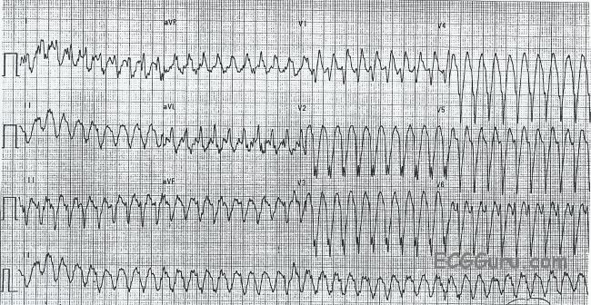
Three types of VT (right or left ventricular outflow tract VT, and fascicular VT) are particularly amenable to treatment by drugs, catheter ablation or both. Specific Noteworthy Ventricular Tachycardias Application of many of the foregoing criteria is shown in Figures 2- -5 5).

16 Further study is needed to determine the true value of this criterion. The algorithm’s performance was substantially less favourable in its first large external application, with an overall accuracy of just 69.0 % compared to, for example, the Brugada criteria (77.5 % accurate) in that same study. The original paper reported remarkable test characteristics with an area under receiver operator curve exceeding 98 %, specificity of 99 % and PPV of 98 %, meaning the algorithm promised to distinguish VT from SVT with 94.5 % accuracy from a single measurement. This is the R wave peak time in lead II, with the interval from QRS onset to first change in polarity (R or S peak) in lead 2 ≥50 ms denoting VT. published the second algorithm offering diagnosis from a single lead, without regard to BBB morphology, and this time using only a single relatively simple measurement. V1 or V2: Onset of r to nadir of S >60 ms → VT Since pre-excited SVTs comprise such a small proportion of WCTs, they will not be considered further in this discussion.

Algorithms have been unable to reliably distinguish between VT and pre-excited SVT, since initial ventricular activation in the latter is (like most VTs) extrinsic to the normal conduction system. As such, most algorithms seeking to discriminate the two entities focus on identifying characteristics unique to VTs – that is to say, those with high specificity for VT – and presume all else to be SVT until proven otherwise the Griffith algorithm is the notable exception to this approach. As a result of this, VT can appear identical to any of the wide complex SVTs above and it is nearly impossible to say with absolute certainty, from an ECG of free-running tachycardia alone, that a wide complex rhythm must be supraventricular. In contrast to the SVTs described above, with relatively constrained modes of ventricular activation, VT can originate from literally any location within the ventricular myocardium or its specialised conduction tissues. Basis and Goals of Ventricular Tachycardia Criteria


 0 kommentar(er)
